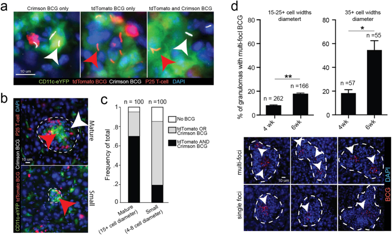
Figure 3. Small DC/P25 T-cell clusters more likely to contain a single BCG strain in mice infected with multiple subtrains.
(A–C) CD11c-eYFP mice were IP infected with a 1:1 (1 × 10⁷ total CFU) mixture of tdTomato BCG and Crimson BCG and 3 weeks later (adoptively transferred (by IV injection) with 5 × 10⁵ DsRed P25 T-cells. Representative areas inside granulomas that contain tdTomato BCG (red arrows) only, Crimson BCG (white arrows) only, or both, in 50 μm-thick liver sections. Low magnification micrographs of the entire granuloma from images in (A) are outlined in white dotted lines in (B) and show two representative sizes of granulomas. (C) Quantification of the frequency of granulomas that contain no BCG, tdTomato or Crimson BCG only, or tdTomato and crimson BCG in 100 mature (15+ cell diameter) and small (4–8 cell diameter) granulomas randomly selected from n = 6 mice. (D) Frequency of granulomas containing multi-focal BCG in 50 μm-thick liver sections in mice 4 or 6 weeks after IP infection with 1 × 10⁷ CFU tdTomato BCG. Representative single and multi-focal granulomas outlined in white dotted lines. White arrows identify individual BCG foci. Granulomas were randomly selected from n = 4 mice in a single experiment. Error bars are mean +/− s.e.m. Student’s t-test used to determine statistical significance. *P < 0.05; **P < 0.01. Number of total granulomas measured for each group shown above individual column.

