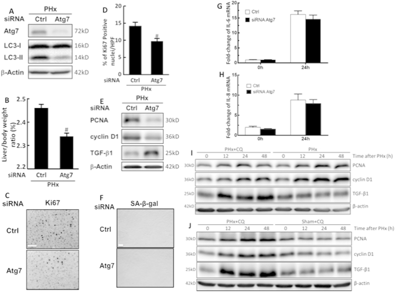
Figure 4. Inhibition of autophagy reduced liver growth and hepatocyte proliferation in the early phase of liver regeneration after PHx.
Wild-type mice were given control or Atg7-specific siRNA for 48 h before treatment with PHx. Liver tissues were harvested at 24 h after surgery and tissue extracts were analyzed for Atg7, LC3-II, and β-actin by Western blotting (A). The liver-to-body-weight ratios were calculated (B). Representative immunohistochemical staining of Ki67 is shown. Scale bar, 50 μm (C). The percentage of Ki67-positive nuclei in hepatocytes was counted in low-power field (200X) in 15 random sections from 3 different mice (D). Tissue extracts were analyzed for PCNA, cyclin D1, TGF-β1, and β-actin by Western blotting (E). Immunohistochemical staining of senescence-associated β-galactosidase (SA-β-gal) in hepatocytes. Scale bar, 100 μm (F). Fold-changes in IL-6 (G) and IL-8 (H) mRNA expression at 24 h after 70% PHx. Wild-type mice were intraperitoneally injected with vehicle (Veh) or chloroquine (CQ) at 0.5 h before PHx or the sham operation and then once per day until 48 h. Liver tissues were harvested at 0–48 h after surgery and tissue extracts were analyzed for PCNA, cyclin D1, TGF-β1, and β-actin by Western blotting (I,J). The values are shown as the mean ± SD in the bar graph and compared using Student’s t test. #P < 0.05 versus control-treated PHx (n = 3).

