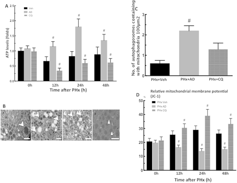
Figure 7. Amiodarone increased removal of damaged mitochondria.
Wild-type mice were intraperitoneally injected with vehicle (Veh), amiodarone (AD), or chloroquine (CQ) at 0.5 h before PHx or sham operation and then once per day until 48 h. Liver tissues were harvested at 0–48 h after surgery. The hepatic ATP concentrations were collected at 0–48 h after PHx (A). Electron microscopic images of autophagosomes containing with mitochondria in PHx mice treated with Veh (a), AD (b), and CQ (c)and the quantification of autophagosomes containing with mitochondria at 24 h after PHx. Arrows indicate autophagosomes containing with mitochondria and arrow heads indicate no structures representing mitochondria itself (d). Scale bar, 2 μm (B,C). Proportion of hepatocytes with a low mitochondrial membrane potential was collected at 0–48 h after PHx (D). The values are shown as the mean ± SD in the bar graph and compared by Student’s t test; #P < 0.05 versus vehicle-treated PHx (n = 6).

