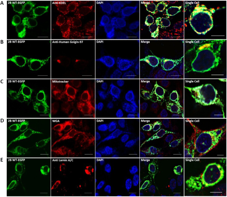
Figure 3. Co-localization of 2B-EGFP with various cellular organelles.
HEK293T cells, transfected with 3 μg 2B-EGFP (green channel) were fixed, permeabilized (for antibody staining), and immunostained with antibodies/dyes (red channel) as follows: (A). Anti-KDEL antibody against ER, (B). Anti-Human Golgin-97 antibody against Golgi bodies, (C). MitoTracker Red FM against mitochondria, (D). Wheat Germ Agglutinin (WGA) Alexa Fluor 594 Conjugate against plasma membrane (E). Anti-Lamin antibody against inner nuclear membrane. Alexa Fluor 555 goat anti-mouse IgG (H+L) was used as secondary antibody and nuclei were counter-stained with 4′,6-diamidino-2-phenylindole (DAPI) dihydrochloride (blue channel). The green, red and blue channels have been merged and shown as a separate panel. The last panel shows the view of a single cell for better visualization of colocalization. Pearson’s correlation coefficient of >0.5 (indicating substantial co-localization) was observed for merged panels of (A,B). The images are representative of cells from at least three areas from three independent experiments. Scale bar - 10 μm, 5 μm (for single cell).

