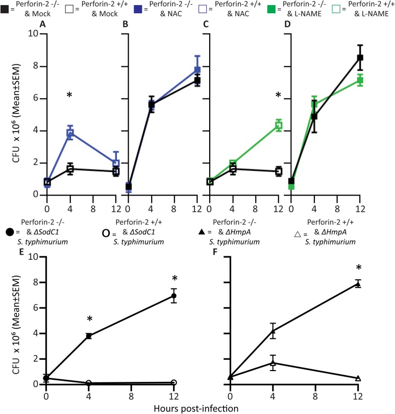
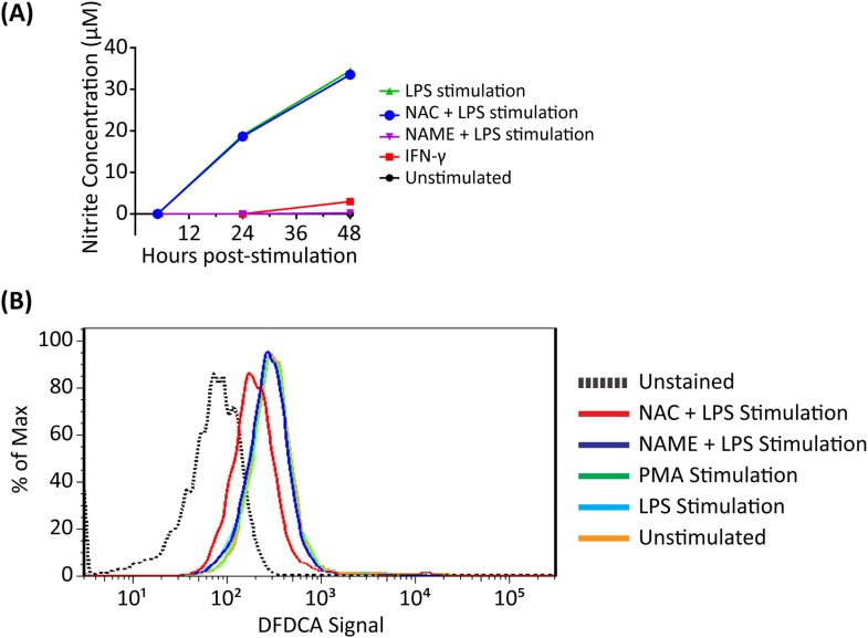
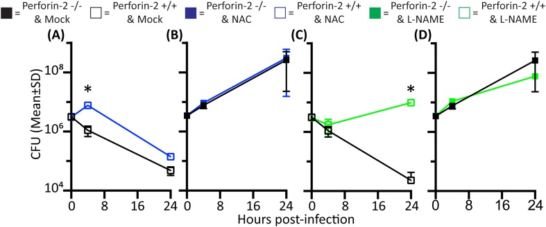
Figure 2. Antimicrobial compounds (ROS and NO) enhance Perforin-2 mediated killing of S. typhimurium by PEM but have limited activity in the absence of Perforin-2.
(A–D) Wild-type S. typhimurium infection of PEMs isolated from either MPEG1 (Perforin-2) +/+ (A, C), or MPEG1 (Perforin-2) −/− mice (B, D). Non-filled symbols indicated MPEG1 (Perforin-2) +/+ PEMs; whereas filled symbols are MPEG1 (Perforin-2) −/− PEMs. Cells were incubated with NAC (blue line), NAME (green line), or mock (black line). To assess bacterial resistance mechanisms against these effectors, (E) SodC1 or (F) HmpA knockout S. typhimurium were used to infect MPEG1 (Perforin-2) −/− or +/+ PEMs. The above experiments were conducted with six biologic replicates and are representative of four independent experiments. Statistical analysis was performed utilizing multiple T-tests with correction for multiple comparisons using the Holm-Sidak method. *p < 0.05.


