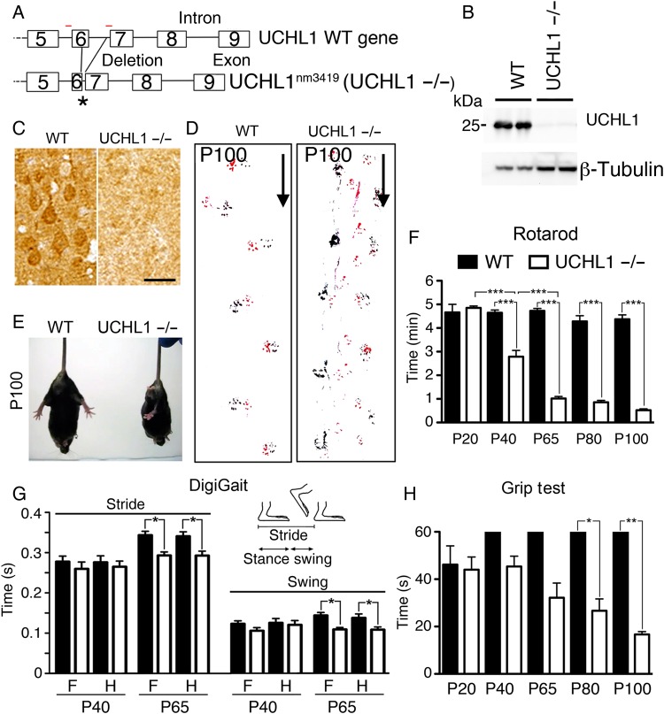
Figure 1.
UCHL1−/− mice show motor function defects. (A) Drawing of Uchl1 gene in WT and UCHL1-deficient (Uchl1nm3419; UCHL1−/−) mice. Partial deletions of exon 6 and intron 6 sequences cause a frameshift and create a de novo stop codon (asterisk). Red lines = location of PCR primers. (B) UCHL1 protein is present in the motor cortex of WT, but not in UCHL1−/−, mice by western blot analysis. β-Tubulin is used as a loading control. (C) UCHL1 expression is present in large pyramidal neurons in the motor cortex of WT, but not in UCHL1−/−, mice. (D) Directional walking patterns of WT and UCHL1−/− mice (marked by arrow) show signs of hindlimb paralysis (red: forelimb; black: hindlimb). (E) Representative screen captures of posture in WT and UCHL1−/− mice tail-hanging at P100. (F) Quantitative analysis of Rotarod test in WT and UCHL1−/− mice at P20 (n = 6 and 6), P40 (n = 14 and 10), P65 (n = 10 and 8), P80 (n = 7 and 9), and P100 (n = 7 and 9). (G) Gait parameter analysis for stride and swing in WT (n = 12) and UCHL1−/− (n = 15) mice at P40 and P65. (H) Quantitative analysis of Grip test in WT and UCHL1−/− mice at P20 (n = 6 and 6), P40 (n = 7 and 12), P65 (n = 7 and 12), P80 (n = 4 and 6), and P100 (n = 3 and 5). Bar graphs represent mean ± SEM. *P < 0.05; **P < 0.01; ***P < 0.001; one-way ANOVA with post hoc Tukey's multiple comparison test. Scale bar, 50 μm.

