Figure 2.
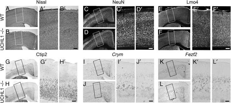
CSMN vulnerability in the UCHL1−/− mice. (A–L) Representative images of matching coronal sections of the motor cortex from WT and UCHL1−/− mice at P100. Nissl staining (A and B), NeuN (C and D), and Lmo4 (E and F) immunocytochemistry in the motor cortex do not show differences between WT and UCHL1−/− mice. However, expression analysis of molecular markers specific to CSMN in layer V of the motor cortex, such as Ctip2 immunocytochemistry (G and H), or Crym (I and J) and Fezf2 (K and L) in situ hybridization show selective reduction of molecular marker expression especially in large pyramidal neurons located in layer V of the motor cortex in UCHL1−/− mice. The boxed areas are enlarged in (A′–L′). Scale bars, 100 μm.

