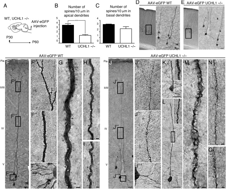
Figure 4.
AAV2-2-mediated retrograde CSMN transduction reveals apical dendrite degeneration and spine loss. (A) Experimental design for AAV2-2-mediated retrograde transduction of CSMN in WT and UCHL1−/− mice. Surgeries are performed at P30 and mice are sacrificed at P60. (B and C) Quantitative analysis of apical and basal dendrites reveals significant reduction in the average numbers of spines in apical (B), but not in basal (C), dendrites. (D–O) Representative images of the motor cortex of WT (D,F–I) and UCHL1−/− (E,J–O) mice. Boxed areas are enlarged to the right. WT CSMN exhibit healthy morphology with a large pyramidal cell body and prominent apical (F′ and F″, G–I) as well as basal dendrites with spines (F‴). CSMN of UCHL1−/− mice show severe cellular degeneration with spine loss throughout the vacuolated apical dendrite (J′ and J″, K–O); but, spines in basal dendrites are not significantly affected (E and J‴). Bar graphs represent mean ± SEM. **P < 0.01, unpaired t-test. Scale bars, (D–F, J) 50 μm, (G–I) and (K–O) 2 μm.

