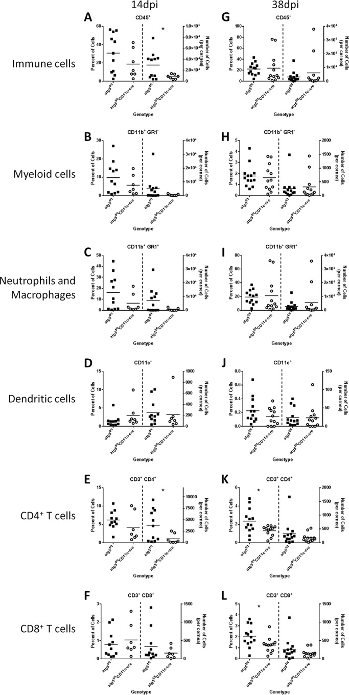
FIG 5 .
DC-autophagy contributes to immune infiltration into the cornea during HSK. atg5fl/fl and atg5fl/fl CD11c-cre female mice were challenged with 2 × 10⁶ PFU of HSV-1 (strain 17) unilaterally. Corneas were analyzed by flow cytometry at 14 dpi (A to F; n ≥ 7 per group) and 38 dpi (G to L; n ≥ 11 per group). Samples were first gated on CD45+ cells before gating on subpopulations. In each panel, quantification of each cell subset is presented as percentage (left) and total number (right) in the cornea. Cell populations analyzed include CD45+ immune cells (A and G), CD11b+ GR1+ neutrophils and macrophages (C and I), CD11b+ GR1− myeloid cells (B and H), CD11c+ dendritic cells (D and J), CD4+ T cells (E and K), and CD8+ T cells (F and L). Statistical significance was determined by unpaired parametric t test. *, P < 0.05.

