
FIG 1 .
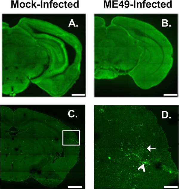
Decreased GABAergic synaptic protein staining in type II ME49 Toxoplasma-infected brains. (A and B) GAD67 immunoreactivity in mock-infected (A) or type II ME49 Toxoplasma-infected (B) brains. (C and D) Detection of type II ME49 Toxoplasma tachyzoites (white arrow in panel D) and bradyzoites (arrowhead in panel D). The image in panel D is a high-magnification image of the boxed in area in panel C. Scale bars, 1 mm (A, B, and C) and 0.25 mm (D). Lines appear in the images due to lack of overlap during image acquisition of each brain.