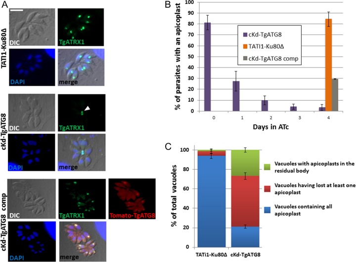
FIG 4 .
TgATG8-depleted tachyzoites rapidly lose apicoplasts, and remaining plastids accumulate in residual bodies. (A) Immunofluorescence assay for detection of apicoplast marker TgATRX1 in HFF monolayers infected with TATi1-Ku80Δ, cKd-TgATG8, and complemented cKd-TgATG8 parasites treated for 4 days with ATc. Tomato-TgATG8 native fluorescence was detected with a Texas Red filter. DNA was labeled with DAPI. The arrowhead indicates apicoplasts within a residual body. Scale bar, 5 µm. (B) Quantification of the number of apicoplasts present in the TgATG8 conditional mutant, parental, and complemented cell lines grown on HFF monolayers for 0, 1, 2, 3, or 4 days in ATc. Values are the mean ± the standard error of the mean of three independent experiments; 200 parasites were counted for each condition. (C) Quantification of the number of vacuoles containing at least one apicoplast within the residual body or with at least one parasite having lost an apicoplast or containing all of their apicoplasts in the cKd-TgATG8 mutant and TATi1-Ku80Δ parental cell lines grown on HFF monolayers for 24 h in ATc. Values are the mean ± the standard error of the mean of two independent experiments, where 200 parasites were counted for each condition.

