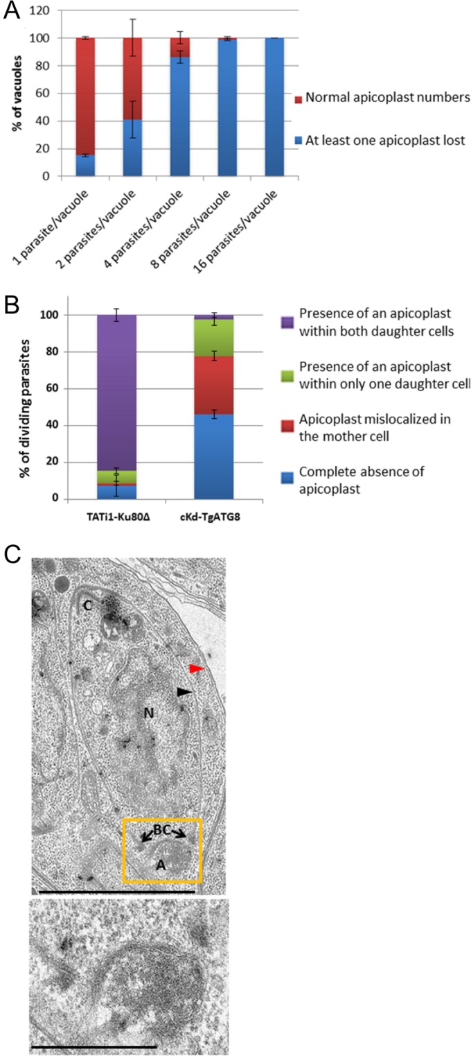
FIG 5 .

Contribution of TgATG8 to apicoplast inheritance. (A) Quantification of the presence of all apicoplasts or the absence of at least one in vacuoles containing 1, 2, 4, 8, or 16 TgATG8 conditional mutant tachyzoites grown on HFF monolayers for 24 h in ATc. Values are the mean ± the standard error of the mean of two independent experiments where 100 parasites were counted for each condition. (B) Quantification of the number of TATi1-Ku80Δ and cKd-TgATG8 dividing tachyzoites incubated for 24 h in ATc that contain no apicoplast or harbor an apicoplast within only one daughter cell or within the mother cells or with apicoplasts normally segregated within both daughters cells. Values are the mean ± the standard error of the mean of two independent experiments, where 200 parasites were counted for each condition. (C) Electron microscopy showing a mislocalized apicoplast outside a forming daughter cell in TgATG8-depleted tachyzoites grown for 24 h in ATc on HFF monolayers. Black arrowhead, daughter IMC; red arrowhead, parent IMC; C, conoid; N, nucleus. The apicoplast (A), of which a magnified view is presented at the bottom, is localized abnormally close to the basal complex (BC). Scale bars, 2 µm (top) and 500 nm (bottom).
