FIG 6 .
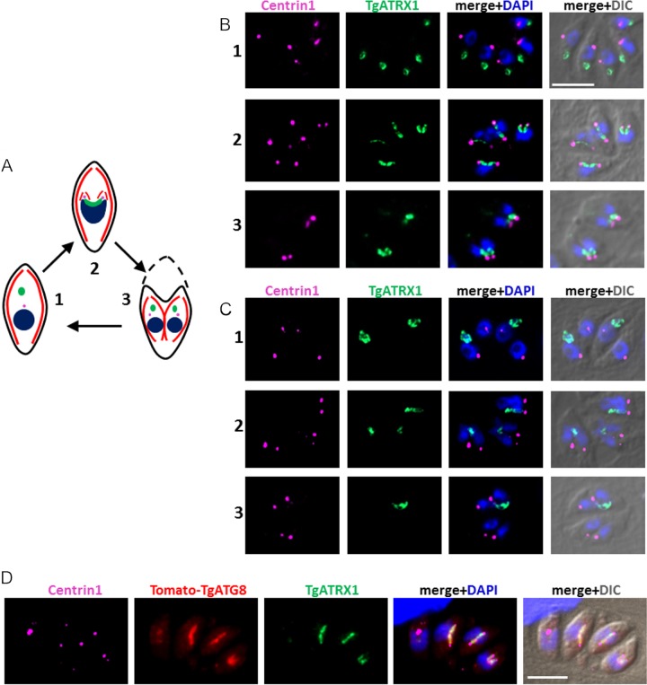
The ends of dividing apicoplasts are no longer linked to centrosomes in TgATG8-depleted tachyzoites. (A) Schematic summary describing apicoplast division and association with centrosomes during endodyogeny. Part 1 shows a mother cell comprising a centrosome and an ellipsoid apicoplast located apical to the nucleus prior to endodyogeny. Part 2 shows that as the nucleus divides, elongated U-shaped apicoplast are associated with the two duplicated centrosomes and inserted into the two growing daughter cells during endodyogeny. Part 3 shows that upon degeneration of the mother cell, fully assembled daughter cells incorporate its plasma membrane at the end of endodyogeny. The apicoplast is green, the nucleus is blue, the centrosome is pink, the IMC is red, and the plasma membrane is black. (B, C) Immunofluorescence analysis of HFF monolayers infected for 24 h in the presence of ATc with parental TATi1-Ku80Δ (B) and mutant cKd-TgATG8 (C) parasites. Different steps of parasite division, corresponding to those described in panel A, are shown. The apicoplast was labeled with anti-TgATRX1 antibody, and centrosomes were labeled with anti-centrin1 antibody. DNA was labeled with DAPI. Scale bar, 5 µm. (D) Immunofluorescence analysis of HFF monolayers infected for 24 h in the presence of ATc with complemented cKd-TgATG8 parasites. The apicoplast was labeled with anti-TgATRX1 antibody, and centrosomes were labeled with anti-centrin1 antibody; Tomato-TgATG8 native fluorescence was detected with a Texas Red filter; DNA was labeled with DAPI. Scale bar, 5 µm. DIC, differential interference contrast.

