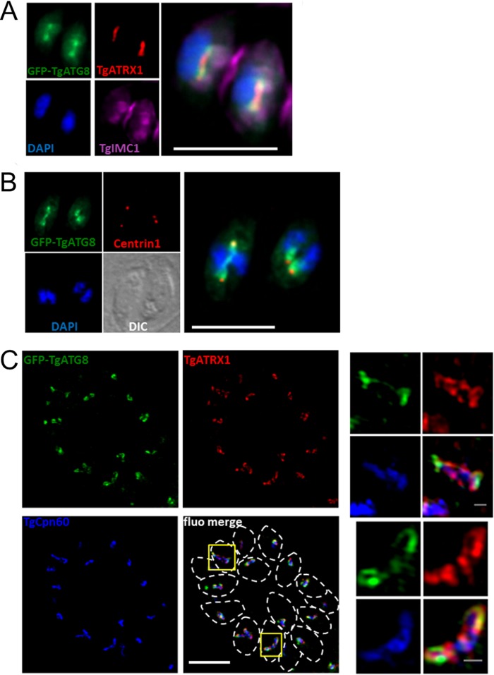
FIG 7 .
TgATG8 is enriched at the ends of elongating apicoplasts. (A) Dividing parasites expressing GFP-TgATG8 with the daughter cell scaffold labeled with TgIMC1 and containing elongated apicoplasts labeled with TgATRX1. DNA was stained with DAPI. A magnified merged image is displayed on the right. (B) In dividing parasites expressing GFP-TgATG8, centrosomes were labeled with anti-centrin1 antibody and DNA was labeled with DAPI. DIC, differential interference contrast. (C) Structured-illumination microscopy imaging of GFP-TgATG8 at elongated apicoplasts together with peripheral membrane protein TgATRX1 and stromal protein TgCPN60. Parasites are delineated by dashed lines in the merged image. Magnified images of selected organelles (yellow squares) are displayed on the right. Scale bars: A and B, 5 µm; C, 5 µm (merged image) and 0.5 µm (magnified images).

