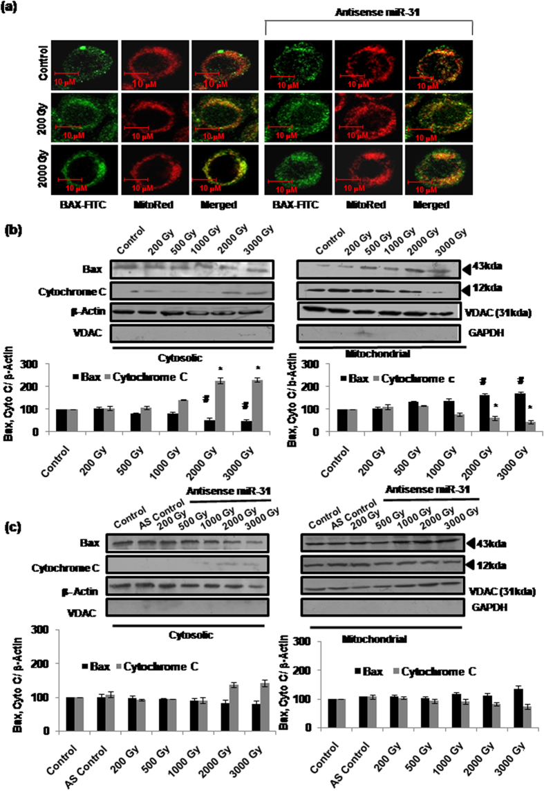
Figure 5. miR-31 dependent translocation of Bax from cytosol to mitochondria corresponds with cytochrome-c release.
(a) Sf9 cells were grown on cover slips and irradiated with or without prior transfection with As-miR-31. Bax protein was tagged using Anti-Bax-FITC antibody conjugate and mitochondria were counterstained with MitoRed. Merged signals confirm the translocation of Bax to mitochondria. Sub-lethal (200Gy) and lethal (2000Gy) radiation doses were used with and without prior treatment with AS-miR-31. Images were captured using 63 × 1.4 NA objective (Leica Sp8 spectral confocal imaging system). Images are representative of four independent experiments. (b) Translocation of Bax to mitochondria (and of cytochrome-c into cytosol) was further assessed by subcellular (cytoplasmic and mitochondrial) fractionation and western immunoblotting at 16 h post-irradiation (#,*P < 0.05). Purity of cytosolic and mitochondrial fractions was confirmed using anti-VDAC and anti- GAPDH antibodies, respectively. (c) Cytosolic and mitochondrial fractions transfected with AS-miR-31 were also used for the western blot analysis for Bax translocation and cytochrome-c release. While β-actin was used as standard loading control for cytosolic fraction, VDAC was taken as loading control for the mitochondrial fraction.

