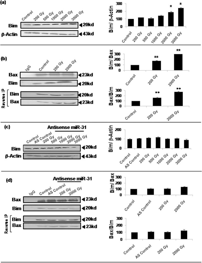
Figure 6. Bim overexpression and its increased interaction with Bax was associated with Bax translocation to mitochondria.
(a) Western blot analysis of Bim was performed 16h post-irradiation for all the selected doses. Densitometric analysis showed a dose-dependent increase in Bim expression (*P < 0.002). (b) Co-immunoprecipitation western immunoblots show highly significant increase in the interaction of Bim with Bax (**P < 0.02). Sf9 cells were irradiated, harvested 16h later, lysed in native condition, followed by immunoprecipitation by anti-Bax antibody. The immunoprecipitated samples were then used for immunodetection of Bim interacting directly with Bax. For further confirmation of Bim –Bax interaction, reverse immunoprecipitation was carried out using anti-Bim antibody and detection by anti-Bax antibody (labeled as ‘Reverse IP’). We chose sub-lethal (200Gy) and lethal (2000Gy) radiation doses for assessing differences in Bim-Bax interaction, and IgG alone was used as antibody control (**P < 0.02). Densitometric analysis was done for the quantitation of all immunoblots. (c,d) Radiation-induced alterations in the expression of Bim as well as its interaction with Bax were also observed after inhibition of Sf-miR-31 by AS-miR-31. Using same radiation doses as above samples were processed for co-immunoprecipitation as well as reverse immunoprecipitation.

