. 2015 Oct 30;4:e09183. doi: 10.7554/eLife.09183

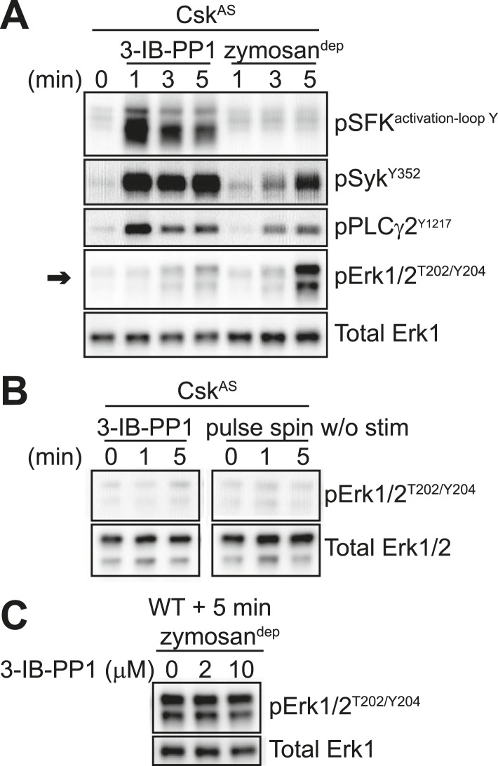
Figure 4. SFK activation in the absence of receptor clustering fails to stimulate downstream signaling in BMDMs.

(A) CskAS BMDMs were pulse-spun with 3-IB-PP1 (10 μM) or with zymosandep (10 particles per cell). Signal transduction was assessed by immunoblotting with antibodies specific to activating phosphorylation sites of the SFKs, Syk, PLCγ2, and Erk (arrow). Total Erk1 is shown as a loading control. (B) CskAS BMDMs were pulse-spun in the presence and absence of 3-IB-PP1, and Erk phosphorylation was assessed by immunoblot. Total Erk1/2 is shown as a loading control. (C) WT BMDMs were treated with zymosandep for 5 min with varying concentrations of 3-IB-PP1 and analyzed by immunoblot. See Figure 4—figure supplement 1 for additional tests of 3-IB-PP1 specificity for CskAS, Figure 4—figure supplement 2 for evidence of that 3-IB-PP1 cotreatment suppresses signaling through the CSF-1 receptor, and Figure 4—figure supplement 3 for evidence that actin-remodeling agents cannot restore 3-IB-PP1-induced downstream signaling.

DOI: http://dx.doi.org/10.7554/eLife.09183.007

Figure 4.

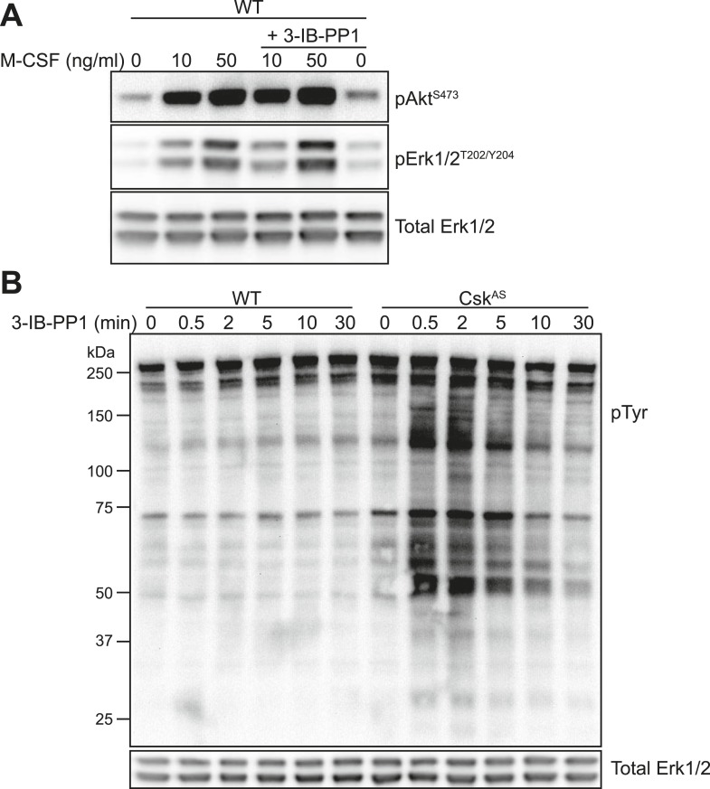
Figure 4—figure supplement 1. Further tests of 3-IB-PP1 specificity for CskAS.

Figure 4—figure supplement 1.

(A) WT BMDMs were treated for 2 min with recombinant M-CSF (10 or 50 ng/ml) in the presence or absence of 3-IB-PP1 (10 µM). Treatment with 3-IB-PP1 does not affect M-CSF-induced phosphorylation of Erk or Akt. (B) WT and CskAS BMDMs were treated to a time course of 3-IB-PP1. Only CskAS cells showed any measurable change in tyrosine phosphorylation (4G10 + pY20 anti-pTyr antibodies).
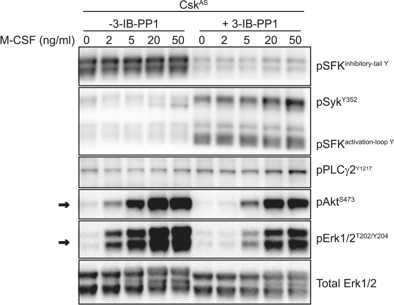
Figure 4—figure supplement 2. Inhibitory effect of 3-IB-PP1 on M-CSF-induced signaling in CskAS cells.

Figure 4—figure supplement 2.

CskAS BMDMs were treated for 5 min with recombinant M-CSF (2, 5, 20, or 50 ng/ml) in the presence or absence of 3-IB-PP1 (10 µM). Addition of 3-IB-PP1 impairs M-CSF induction of Akt and Erk phosphorylation (arrows), suggesting an active, inhibitory effect of 3-IB-PP1 treatment that blocks downstream signaling.
Figure 4—figure supplement 3. Actin-remodeling agents do not restore signaling downstream of 3-IB-PP1 treatment.

Figure 4—figure supplement 3.

BMDMs were stimulated with 3-IB-PP1 or zymosandep in combination with the actin-remodeling agents Cytochalasin D (CytD, 10 µM), Latrunculin A (LtrA, 0.5 µM), and Jasplakinolide (Jas, 1 µM). Although each compound affected zymosandep signaling, none restored Erk phosphorylation after 3-IB-PP1 treatment (CytD n = 2). Note that these images were developed with high signal, so the basal Erk phosphorylation looks high even though the cells have not been activated. The important point is that there is no increase in Erk phosphorylation with 3-IB-PP1 treatment.