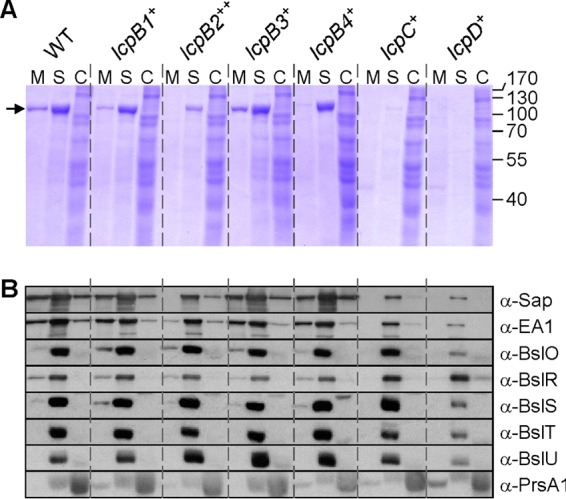
FIG 3.

Subcellular fractionation of proteins with SLH domains. (A) Cultures of wild-type (WT) B. anthracis, carrying all six lcp genes or of variants with a single lcp gene (lcpB1+, lcpB2++, lcpB3+, lcpB4+, lcpC+, or lcpD+) were grown to an A600 of 2. Cultures were centrifuged to separate the extracellular medium (M) from the bacterial sediment. Proteins in the S layers (S) were extracted by boiling cells in 3 M urea. Stripped cells were broken mechanically to release cytosolic extracts (C). Proteins in all fractions were precipitated with TCA, washed in acetone, and separated by 10% SDS-PAGE. The gel was stained with Coomassie brilliant blue. The positions of Sap and EA1 that migrate with the same mobility on SDS-PAGE and of molecular mass markers are indicated by an arrow, and masses are given in kilodaltons on the right. (B) B. anthracis cultures fractionated as described for panel A were subjected to immunoblotting with rabbit antisera raised against purified Sap, EA1, BslO, BslR, BslS, BslT, BslU, or PrsA1.
