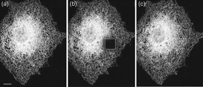
Figure 3. Photobleaching.

Cells expressing a GFP-labelled protein in the endoplasmic reticulum were subjected to photobleaching. (a) A cell before bleaching. (b) The same cell immediately after bleaching of the square section shown. (c) The same cell 5 min after photobleaching. Adapted from Figure 1b from Lippincott-Schwartz, J., Snapp, E. and Kenworthy, A. (2001) Studying protein dynamics in living cells. Nat. Rev. Mol. Cell. Biol. 2, 444–456.
