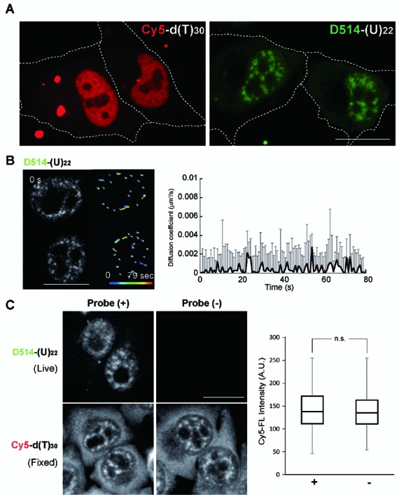
Figure 2.

ECHO-liveFISH imaging of poly(A) RNA foci in living HeLa cells reveals immobility. (A) Fluorescence images of HeLa cells transfected with Cy5-d(T)30 or D514-(U)22. Speckled nuclear fluorescence was readily distinguished in the nuclei of D514-(U)22 transfected cells but not in the Cy5-d(T)30 transfected cells. (B) Time-lapse confocal poly(A) RNA images (LSM780) of HeLa cells transfected with D514-(U)22 (also see Supplementary Video 1). The track-line presentations monitor the center position of individual speckles over time (progressing from blue to red). Diffusion coefficient of each poly(A) speckle was plotted over time. (C) Poly(A) RNA staining in HeLa cells after ECHO-liveFISH imaging. Top, D514-(U)22 fluorescence in transfected and mock-transfected cells. Bottom, after imaging with D514-(U)22, cells were fixed, permeabilized and hybridized with Cy5-d(T)30 probes followed by Cy5 fluorescence imaging. Quantification of mean Cy5 fluorescence intensity at individual speckles (MFI) indicated comparable endogenous poly(A) concentrations at nuclear foci in D514-(U)22-transfected and mock-transfected cells. Scale bars: 20 μm. D514: 514 nm excitation, 517–597 nm detection; Cy5: 633 nm excitation, 638–759 nm detection. 0.264 × 0.264 μm pixel size; 50 μs pixel dwell time.
