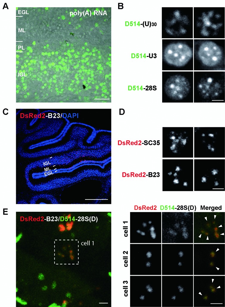
Figure 6.

ECHO-liveFISH imaging of target RNA intranuclear foci in acute mouse brain tissues. (A) A representative confocal image from acute cerebellar slices prepared soon after in vivo electroporation. (B) Confocal images (FV1000) of poly(A), U3 snoRNA and 28S rRNA in individual cerebellar granule cells after electroporation. Intranuclear foci containing target RNA concentrations are readily distinguished. The number and shape are consistent with foci of nuclear speckles and nucleoli. (C) A confocal image of a DsRed2-B23 electroporated mouse cerebellum stained with a DsRed2-specific antibody (red) and DAPI (blue). (D) High magnification confocal images of DsRed2-SC35 and DsRed2-B23 in individual nuclei. Note the irregular shapes of DsRed2-SC35 speckles and round-shaped DsRed2-B23 foci. (E) Colocalization between DsRed2-B23 and D514-28S at the nucleoli of electroporated granule neurons. P10 mice expressing DsRed2-B23 DNA plasmids were perfused and cerebellar slices were processed for DsRed2 and DAPI staining simultaneously with D514-28S hybridization. Overlapping DsRed2 and D514 fluorescence at nuclear foci indicates colocalization between B23 proteins and 28S rRNA (white arrowheads). EGL: external granule layer, ML: molecular layer, PL: Purkinje layer, IGL: inner granule layer. Scale bars: 20 μm (A), 2.5 μm (B, C2, D) and 1 mm (C1). DAPI: 405 nm excitation, 425–475 nm detection; D514: 515 nm excitation, 530–575 nm detection; DsRed2: 561 nm excitation, 566–703 nm detection. 0.207 × 0.207 μm pixel size; 8 μs pixel dwell times.
