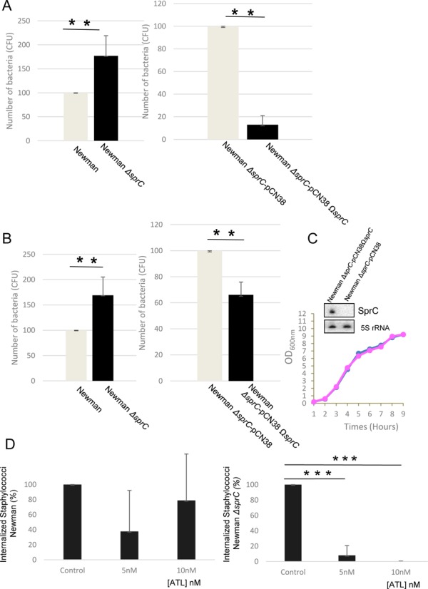
Figure 5.

SprC reduces S. aureus internalization by human macrophages and its absence augments phagocytosis and bacterial release from the host cells. (A) Number of S. aureus cells (CFU) after internalization. Newman, Newman-ΔsprC, Newman-ΔsprC pCN38 and Newman-ΔsprC pCN38ΩsprC cells were incubated with the THP1 macrophages (MOI 1:25) for 2 h. The non-phagocytosed bacteria were killed by culturing in medium containing 50 μg/ml gentamycin for 24 h. The medium was then replaced with fresh media for up to 4 days. (B) After 4 days, colony counts correspond to the bacteria that were released by the human macrophages. The data presented are the mean ± SD of three independent experiments realized in triplicates. The data are considered highly significant for P-values ≤0.01 (**). (C) Monitoring bacterial growth and SprC expression levels, by Northern blots (OD600nm = 2), in S. aureus strains Newman ΔsprC-pCN38 (pink) and Newman ΔsprC-pCN38ΩsprC (blue). The 5S rRNAs are the loading controls. Each blot presented is from identical experiments. (D) Inhibition of S. aureus strain Newman ΔsprC phagocytosis by the THP1 macrophages by adding increasing concentrations of purified ATL protein (right panel) whereas exogenous ATL has limited, if any, effects on S. aureus strain Newman internalization (left panel).
