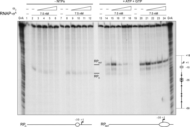
Figure 4.

Effect of ω2 on the formation of RPO at Pω. The 423-bp [α32P]-Pω DNA (1 nM) was pre-incubated with increasing concentrations of ω2 (7.5, 15, 30 and 60 nM; lanes 2–6 and 14–18) or with 7.5 nM RNAP-σA (lanes 8–12 and 20–24) in buffer C. A second protein was added along with the initiating nucleotides, GTP and ATP (as indicated). DNA melting was probed by KMnO4 footprinting as a way of observing the open complex. The positions hypersensitive to KMnO4 are labelled (RPO and RPINIT) and depicted at the bottom of the figure. The coordinates are relative to the transcription start point. Chemical sequencing reactions for purines (G +A) are shown and the relevant regions of Pω depicted to the right of the figure.
