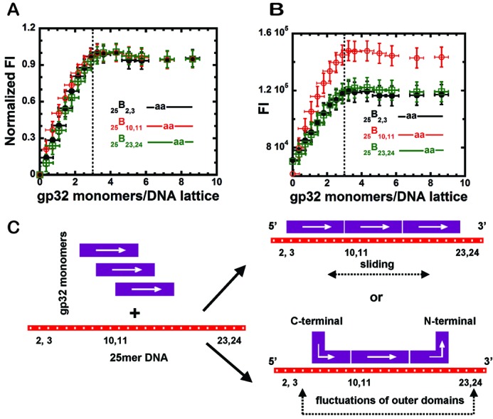
Figure 1.

Fluorescence intensity changes at 370 nm observed for gp32 binding to 25-mer ssDNA constructs with 2-AP dimer probes located at different positions, and a schematic model for the binding. (A) Normalized fluorescence intensity changes of the dimer probes for ssDNA constructs at 1 μM concentration as a function of gp32 monomers added per 25-mer ssDNA lattice. 25B2,3 construct (black, closed circles); 25B10,11 construct (red, open circles) and 25B23,24 construct (green, open squares). The dashed vertical line in (B) represents the point at which 3.0 gp32 monomers have been added per 25-mer ssDNA lattice. (B) Raw fluorescence intensity changes for the titration curves of the same ssDNA constructs as in Figure 1A (same color-coding). (C) Schematic model of possible binding modes of trimeric gp32 monomer clusters to these 25mer ssDNA lattices.
