Figure 6.
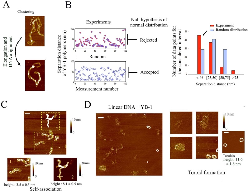
YB-1 multimerization at DNA crosses leads to the alignment of the two interacting DNA helices and higher-order assembly of DNA molecules. (A) Clusters of YB-1 (300 nM) multimers are clearly detected on supercoiled DNA (2 nM) and are especially located at DNA crosses (upper image). When many YB-1 multimers are bound to supercoiled DNA, the molecule is forced to adopt a linear shape (lower image). Scale bar: 50 nm. (B) When the trajectory adopted by DNA helices is clearly distinguished by AFM all along supercoiled DNA, we measured the separation distance between two consecutive YB-1 multimers along DNA. We then plotted a theoretical curve of a random distribution using the same number of data and the same length of DNA. Separation distances smaller than 10 nm were discarded to mimic the limit imposed by the AFM resolution. We then tested the null hypothesis of a normal distribution for these two cases using the Chi-square goodness-of-fit test at the 5% significance level. In contrast with a random distribution, the null hypothesis was rejected for the experimental results. In agreement with this, the bar plot indicates a higher occurrence of YB-1 which are close from each other (<25 nm) (see arrow). (C) Supercoiled DNA (2 nM) were assembled into high order structures on mica in the presence of YB-1 at high concentration (1 μM), as observed by AFM. In the lower right panel, the height of the nucleoprotein structure of cylindrical shape indicates that at least two supercoiled DNA are bundled. The lower left panel shows an intermediary structure of DNA bundles with two supercoiled DNA which are self-aligned. Scale bar: 150 nm. (D) YB-1 at high concentration (1 μM) leads to the alignment of 1220 bp linear DNA into large circular structure on mica which, at the end of the process, formed perfect toroids, as observed by AFM. The height of the toroids indicates that many DNA are bundled. Interestingly, free 1220 bp linear DNA coexists with toroids on mica, which suggests a cooperative binding of YB-1 to DNA. Scale bars: 150 nm.

