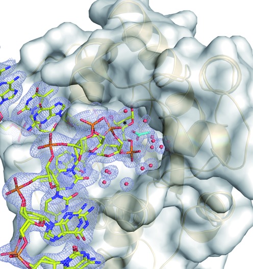
Figure 5.

The new structures reveal a solvent-filled channel to the active site for DNA-bound TDG. The structure of the enzyme–product complex resulting from TDGcat action on a G·U DNA substrate (PDB ID: 4Z47, 1.45 Å) reveals a solvent-filled channel from the active site to the enzyme surface that runs along the target DNA strand. TDGcat is shown in both space-filling and cartoon modes, the DNA is in stick format, with the target strand colored yellow (complementary strand not shown for clarity). Water molecules are shown as red spheres, and the acetate is cyan. The 2Fo-Fc omit map, contoured at 1.0 σ, is shown light blue for the target DNA strand, acetate and water molecules.
