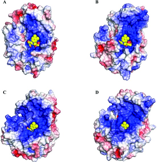
Figure 5.

Comparison of the electrostatic surface of the TIM-barrel and helical domains of HsDus2 (A), DUS from T.thermophilus (B, pdb 3b0p), from E.coli (C, pdb 4bfa) and from a putative flavin oxidoreductase from T.maritima (D, pdb 1vhn). The electrostatic potentials displayed are within ± 5 kTe−1 and were calculated with APBS. The electrostatic surface of the helical domain of HsDus2 is less positively charged with respect to the bacterial dihydrouridine synthases and may explain the weak interaction of HsDus2dusD with tRNA. The FMN is shown in yellow spheres.
