
Figure 4. miR-23b suppresses EMT and sphere formation ability of gastric cancer cells.
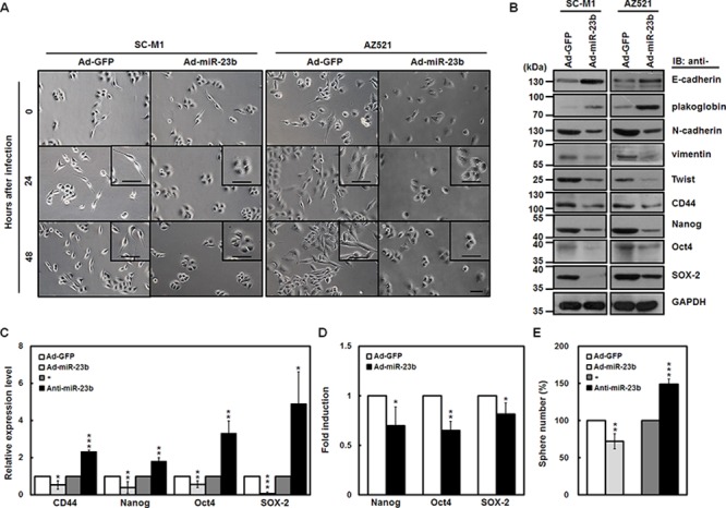
A. SC-M1 (left) and AZ521 (right) cells were infected with adenoviruses expressing miR-23b (Ad-miR-23b) or GFP (Ad-GFP) and subsequently seeded onto 6-well plates for 24 or 48 hours for morphological examination. Bar, 50 μm. B. Whole-cell extracts of the infected SC-M1 (left) and AZ521 (right) cells were prepared for Western blot analysis using anti-E-cadherin, anti-plakoglobin, anti-N-cadherin, anti-vimentin, anti-Twist, anti-CD44, anti-Nanog, anti-Oct4, anti-SOX-2, and anti-GAPDH antibodies. C. After infection with adenoviruses expressing miR-23b or GFP or transfection with 100 nM antagomir-23b (anti-miR-23b) or scrambled control (−) into SC-M1 cells, the transcript levels of CD44, Nanog, Oct4, and SOX-2 were measured by quantitative real-time PCR and then normalized to GAPDH. *P < 0.05; **P < 0.01; ***P < 0.001. D. After transfection with reporter plasmids Nanog-Luc (Nanog), Oct4-Luc (Oct4), or SOX-2-Luc (SOX-2) for 24 hours, SC-M1 cells were infected with adenoviruses expressing miR-23b or GFP for 24 hours for reporter gene assay. *P < 0.05; **P < 0.01. E. After infection with adenoviruses expressing miR-23b or GFP or transfection with 100 nM antagomir-23b or scrambled control into SC-M1 cells for 48 hours, the treated cells were seeded and then incubated for 9 days for tumorsphere formation assay. **P < 0.01; ***P < 0.001. Data are shown as mean ± standard deviation.