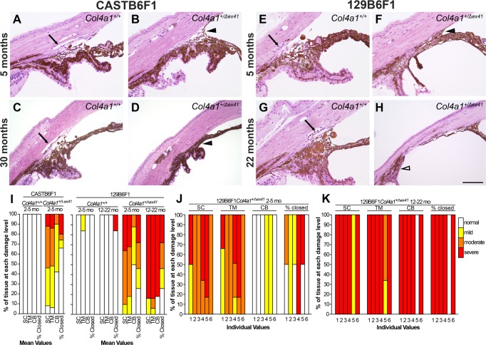
Figure 2.
Histologic analyses of iridocorneal angle dysgenesis. Histologic analyses revealed morphologic defects of the iridocorneal angle that progressed with age. In control eyes (A, C, E, G) there was a robust, foliated ciliary body, clearly identifiable Schlemm's canal (arrow), and trabecular meshwork. Analyses of Col4a1+/Δex41 CASTB6F1 mice (B, D) showed prevalent, but usually mild, iridocorneal angle defects with hypoplastic or absent Schlemm's canal (13/15 eyes), hypoplastic trabecular meshwork (13/15 eyes), and anterior synechia (10/15 eyes; black arrowhead). Frequencies are indicated for 5-month-old mice. Eyes from Col4a1+/Δex41 mice on the 129B6F1 background between 1.5 and 5 months of age (F) showed hypoplastic Schlemm's canal, trabecular meshwork, and ciliary body as well as anterior synechia (black arrowhead; n = 6). Eyes from mice between 22 and 31 months (H) had severe and extensive iridocorneal adhesions and an unrecognizable Schlemm's canal and trabecular meshwork (n = 6). The pars plana portion of the ciliary body is indicated (white arrowhead). Semiquantitative analyses of iridocorneal dysgenesis for each strain or for individual Col4a1+/Δex41 129B6F1 eyes are shown in ([I–K], respectively). Normal Schlemm's canal (SC), trabecular meshwork (TM), and ciliary body (CB) are indicated with white bars. Mild pathology (yellow) indicates that 50% to 100% of the TM, SC, or CB is present; moderate pathology (orange) indicates that 10% to 49% of these regions are present and severe (red) indicates that less than 10% of these regions are present. The percent angle closure was scored as normal (0%–25% closed), mild (25%–50% closed), moderate (50%–75% closed), or severe (>75% closed). Scale bar: 100 μm.

