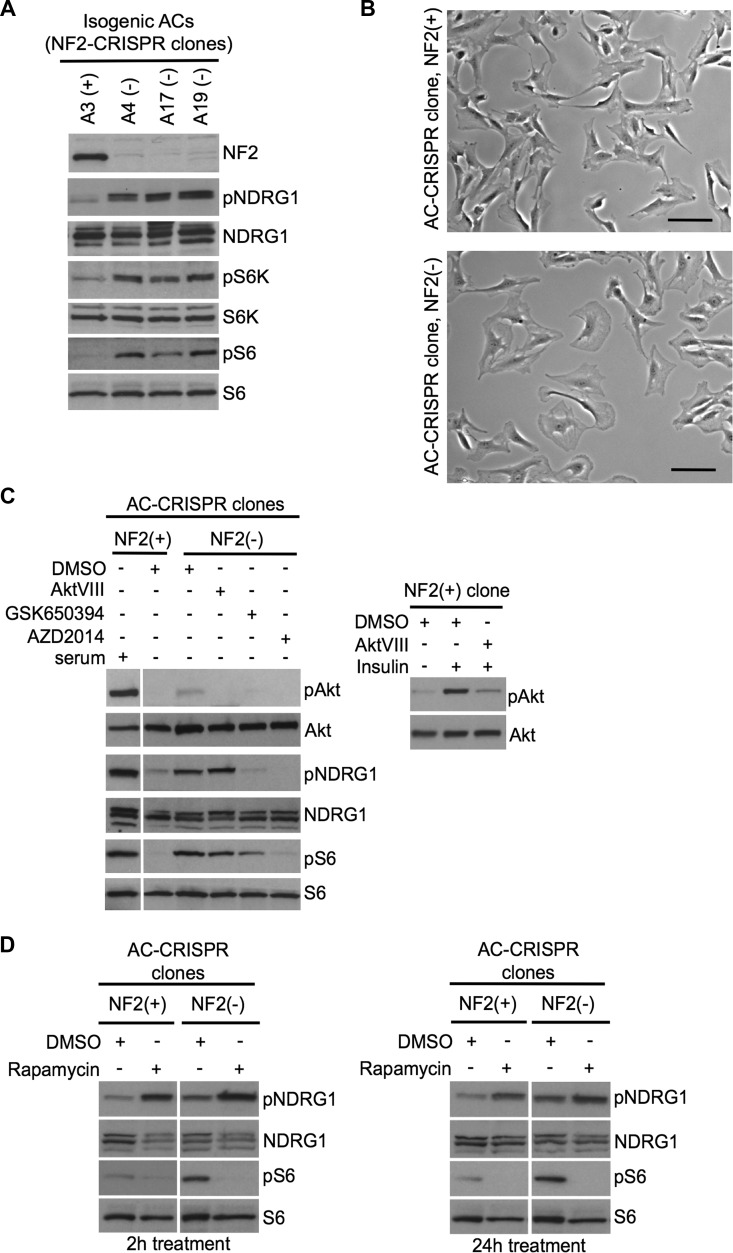
Figure 5. Inactivation of NF2 using CRISPR-Cas9 genome editing in human arachnoidal cells recapitulates enlarged cell morphology and signaling signatures of NF2-deficient human meningioma cells.
A. Immunoblotting with indicated antibodies of NF2-null (−) AC-CRISPR clone compared with NF2-expressing (+) AC-CRISPR clones demonstrates loss of NF2 expression, increased p70S6K (T389) phosphorylation (pS6K) and pS6 (mTORC1 readouts), and increased pNDRG1 (SGK1 readout) compared with NF2-expressing (+) clone. S6K, S6 and NDRG1 serve as controls. B. Representative bright-field images show enlarged cell morphology in NF2-null (A17) AC-CRISPR clone (right) compared to NF2-expressing (A3) clone (left). Scale bar = 100 μm. C. Left panel shows immunoblot analysis with indicated antibodies of an NF2(−) compared to NF2(+) AC-CRISPR clones treated with GSK650394 (2μM, 18h), dual mTOC1/2 kinase inhibitor AZD2014 (300 nM, 2h), AktVIII (1 μM, 2h) inhibitors or DMSO alone. As a control for Akt inhibition, AktVIII treatment of insulin-stimulated NF2(+) AC-CRISPR clone is shown (right panel). D. Immunoblotting with indicated antibodies of NF2(+) and NF2(−) AC-CRISPR clones treated with rapamycin (20nM) or DMSO for 2h (left panel) and 24h (right panel) shows an increase in pNDRG1 after rapamycin treatment.

