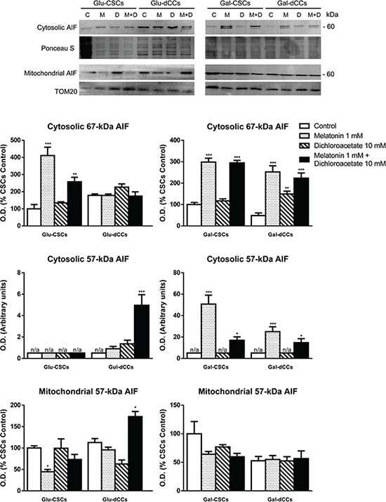
Figure 5. Representative immunoblot images for apoptosis-inducing factor (AIF) in cytosolic and mitochondrial extracts from P19 stem (CSCs) and differentiated (dCCs) cells, grown in glucose (Glu) and galactose (Gal) media, show a higher cytosolic localization of ~57 kDa form of AIF in cells grown in galactose media, treated with melatonin, alone or in combination with dichloroacetate.

Treatments: control (C), melatonin 1 mM (M), dichloroacetate 10 mM (D), and melatonin 1 mM + dichloroacetate 10 mM (M+D). Ponceau S and TOM20 were used to normalize sample loading in cytosolic and mitochondrial extracts, respectively. Bar charts show means of normalized optical density (O.D.) ± SEM, from at least three separate immunoblots. Statistical comparisons: *p < 0.05; **p < 0.01; ***p < 0.001 vs. control. n/a: no band detected (O.D. = 0).
