
Figure 1. Protoporphyrin IX accumulation in PECA cells.
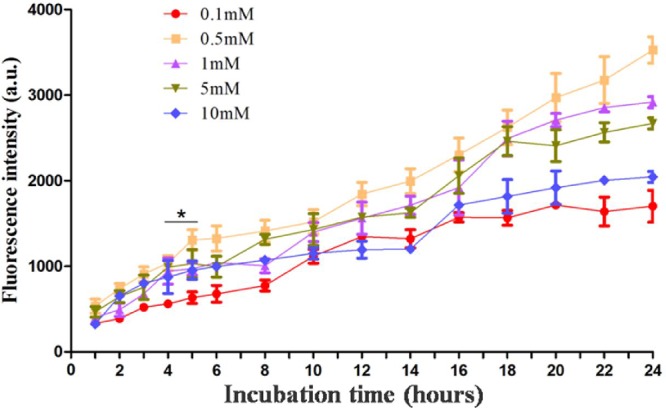
PECA cells were incubated with ALA of different concentrations (0.1 to 10 mM) in serum-free medium in the dark with different incubation times (1 to 24 h). PpIX fluorescence emission from the cells was detected using a microplate reader (n = 5). PpIX accumulation in PECA cells increased with the incubation time. PpIX fluorescence in cells incubated with 0.5 mM ALA was higher than that from cells incubated with other ALA concentrations. PpIX accumulation in PECA cells incubated with 0.5 mM ALA for 5 h was higher than that for 4 h (p < 0.05), but close to that for 6 h (p > 0.05).