
Figure 6. Secretion of IL-12 and IL-10 from DCs.
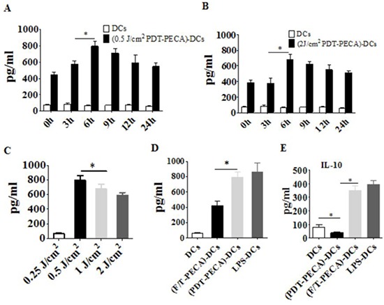
Secretion of IL-12 from DCs after incubation with PECA cells treated by ALA-PDT with 630 nm LED light at doses of 0.5 J/cm2 A. or 2 J/cm2 B. at different times (0 to 24 h) after light irradiation, with unstimulated imDCs as a control. C. PECA cells were treated by ALA-PDT with different light doses (0.25 to 2 J/cm2). Six hours after treatment, PECA cells were collected and incubated with imDCs for 24 h and IL-12 secreted from the DCs were quantified. D. and E. PECA cells treated by PDT (0.5 mM ALA, 0.5 J/cm2) or three cycles of freeze thaw (F/T). Six hours after treatment, PECA cells were collected and incubated with imDCs for 24 h and IL-12 (D) or IL-10 (E) was measured. Secretion of IL-12 or IL-10 form unstimulated imDCs was used as a negative control and secretion of IL-12 or IL-10 form DCs after incubation for 24 h with LPS was used as a positive control.