
Figure 7. T cell proliferation stimulated by DCs.
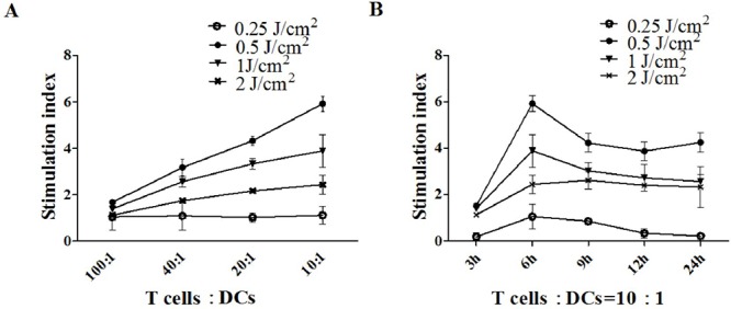
A. PECA cells were treated by ALA-PDT with different light doses (0.25 to 2 J/cm2). Six hours after treatment, PECA cells were incubated with imDCs for 24 h. Then T cells were incubated with collected DCs in different ratios (100:1, 40:1, 20:1, 10:1) for 72 h (n = 5). B. PECA cells were treated by PDT with different light doses (0.25 to 2 J/cm2). At different times (3 to 24 h) after PDT, PECA cells were incubated with imDCs for 24 h. Then T cells were incubated with collected DCs at a ratio of 10:1. Untreated T cell were used for a negative control, T cell proliferation was quantified by CCK-8 assay. ALA-PDT-treated PECA cells enhanced the capability of DCs to induce T cell proliferation. DCs stimulated by PDT induced apoptotic PECA cells are more potent stimulators of T cell proliferation than DCs stimulated by PDT induced necrotic PECA cells.