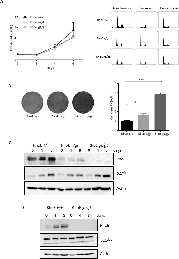
Figure 1. RhoE is a mediator of contact inhibition.

A. Lack of RhoE expression does not affect cell proliferation. Left: Primary MEFs were grown in DMEM-10% FBS and fixed at the indicated time points. Cell density was measured by crystal violet staining. Data (referred to time 0) from three independent experiments are shown as Mean+SEM. A.U.: arbitrary units. Right: Cell cycle profile of primary MEFs growing in 10% FBS (Asynchronous), serum-starved for 48 h (No serum) or re-stimulated for 16 h after serum-starvation (Re-stimulated) was analyzed by flow cytometry after DNA staining with Propidium Iodide. B. RhoE deficient cells are not contact inhibited. RhoE+/+, RhoE+/gt and RhoEgt/gt primary MEFs were kept in culture for 15 days and cell density was measured by crystal violet staining. Pictures show examples of the plates after staining. The graph shows the Mean+SEM of three independent experiments (*p < 0.05 and ***p < 0.001 in a Student's t test). A.U.: arbitrary units. C. p27Kip1 accumulates normally in high density cultures in the absence of RhoE. Primary MEFs as in B were kept in culture for 8 days with medium-change every 48 h. At the indicated time-points, the expression of RhoE and p27Kip1 was analyzed by Western blotting. Actin was used as a loading control. D. p21Cip1 expression does not change in the absence of RhoE expression. RhoE+/+ and RhoEgt/gt primary MEFs were kept in culture for 8 days and the expression of p21Cip1 and RhoE was analyzed as in C.
