
Figure 4. RhoE protects from DMBA/TPA-induced skin tumors in mice.
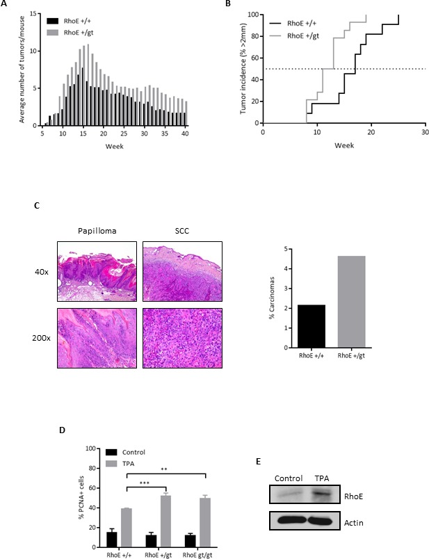
A. Average number of tumors in RhoE+/+ and RhoE+/gt mice (n = 14 each) treated with DMBA/TPA. TPA treatment was discontinued after week 15. Differences between both genotypes were significant in a 2 way ANOVA (p < 0.0001). B. Percentage of mice having tumors bigger than 2 mm. p = 0.0145 in a Mantel-Cox test. C. After the DMBA/TPA treatment, mice were sacrificed and lesions classified as papilloma or squamous cell carcinoma (SCC). Representative images of papilloma (left) and SCC (right) from a RhoE+/gt mouse are shown. Percentage of carcinomas relative to the maximum number of papillomas is represented (right). In RhoE+/+ mice, 1 carcinoma was found from a maximum of 47 papillomas at week 15, whereas in the case of RhoE+/gt mice, 7 carcinomas from 151 papillomas were found. Thus, the conversion rate from papilloma to carcinoma was 2.1% for RhoE+/+ and 4.6% for RhoE+/gt mice, and is shown in the graph on the right. D. RhoE expression reduces the induction of proliferation by TPA. Mice of the three genotypes (RhoE+/+, RhoE+/gt and RhoEgt/gt, n = 3 of each one) were treated with a single dose of TPA (12.5 μg in 0.2 ml acetone) for 24 h. Percentage of PCNA positive nuclei determined by immunohistochemistry is plotted (**p < 0.01 and ***p < 0.001 in a 2 way ANOVA followed by Bonferroni's multiple comparisons test). E. TPA induces the expression of RhoE in skin. RhoE expression in skin samples from control and TPA-treated RhoE+/+ mice (as in A) was analyzed by Western blotting. Actin was used as a loading control.