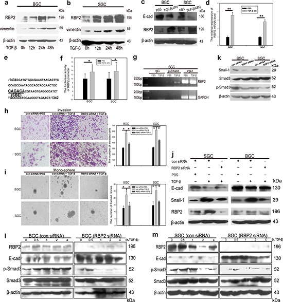
Figure 3. RBP2 can be induced by TGF-β1 and RBP2 is essential for EMT induced by TGF-β1.

a. and b. TGF-β1 induces RBP2 protein expression in a time dependent manner in BGC-823 and SGC-7901 cells respectively. Representative images are shown here from three independent biological replicates. c. Upregulation of RBP2 and downregulation of E-cadherin by TGF-β1 treatment for 48 hours using western blot. Representative images are shown here from three independent biological replicates. d. QRT-PCR shows induction of RBP2 mRNA level in GC cell lines by TGF-β1 treatment for 48 hours. Data are mean ± SD of 3 biological replicates, **p < 0.01 compared with negative control. e. SBE element in RBP2 promoter. f. Increase of promoter luciferase activity of RBP2 by TGF-β1 treatment for 12 hours. Data are mean ± SD of 3 biological replicates, *p < 0.05 compared with negative control. g. p-Smad3 directly binds to RBP2 promoter using ChIP assay. h. and i. Invasion and maintenance of stem cell property of GC cells induced by TGF-β1 can be abrogated with RBP2 inhibition respectively. Data are mean±SD of 3 biological replicates, *p < 0.05 compared with negative control. Original magnification, × 60 and × 40 respectively. j. RBP2 suppression reverted induction of mesenchymal markers and inhibition of epithelial marker mediated by TGF-β1. Representative images are shown here from three independent biological replicates. k. Inhibition of RBP2 decreases endogenous Smad3 phosphorylation. Representative images are shown here from three independent biological replicates. l. and m. Pre-treatment with RBP2 siRNA in GC cell lines markedly retrieves Smad3 activation (formation of p-Smad3) induced by TGF-β1 in different time points in BGC-823 and SGC-7901 cells respectively.
