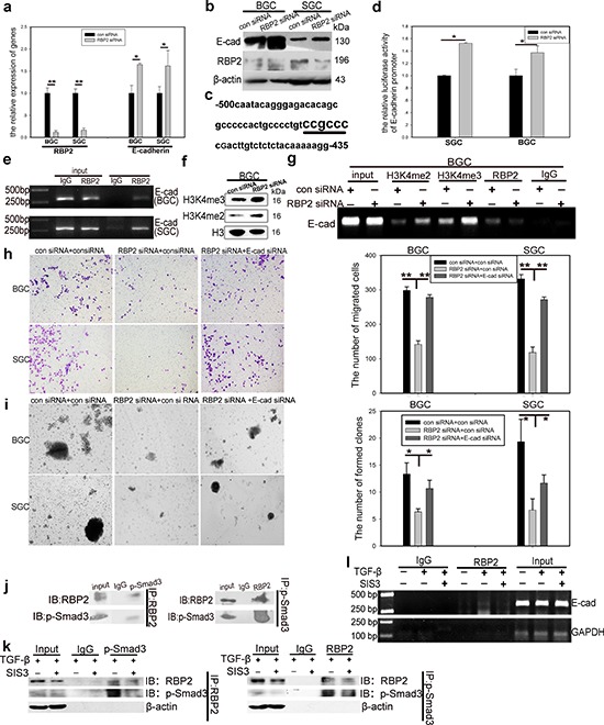
Figure 4. RBP2 participates in the regulation of GC progression by directly suppressing E-cadherin expression and p-smad3 binds to RBP2 and recruits it to the promoter of E-cadherin to reinforce suppression effect.

a. and b. QRT-PCR and western blot results show downregulation of RBP2 and upregulation of E-cadherin in GC cells with RBP2 inhibition. Data are mean ± SD of 3 biological replicates, * and **p < 0.05 and < 0.01 compared with negative control. c. RBP2 recognizing element CCGCCC in E-cadherin promoter. d. E-cadherin promoter luciferase activity increases when RBP2 is depleted in GC cells. Data are mean ± SD of 3 biological replicates, *p < 0.05 compared with negative control. e. ChIP assay shows direct binding of RBP2 to E-cadherin promoter. f. The levels of H3K4me2 and H3K4me3 increase in GC cells with RBP2 inhibition. g. Binding of RBP2 to E-cadherin promoter is its histone demethylase dependent. h. and i. Decrease of migration and maintenance of stem cell property of GC cells induced by RBP2 inhibition is abrogated by E-cadherin suppression simultaneously. Data are mean±SD of 3 biological replicates, * and **p < 0.05 and < 0.01 compared with negative control. Original magnification, × 40. j. Immunoprecipitate assay indicates the interaction between p-smad3 and RBP2. Representative images are shown here from three independent biological replicates. k. Immunoprecipitate assay demonstrates SIS3 decreases RBP2 induction by TGF-β1 and diminishes the interaction between p-smad3 and RBP2. l. Binding of RBP2 to E-cadherin promoter decreases with SIS3 treatment.
