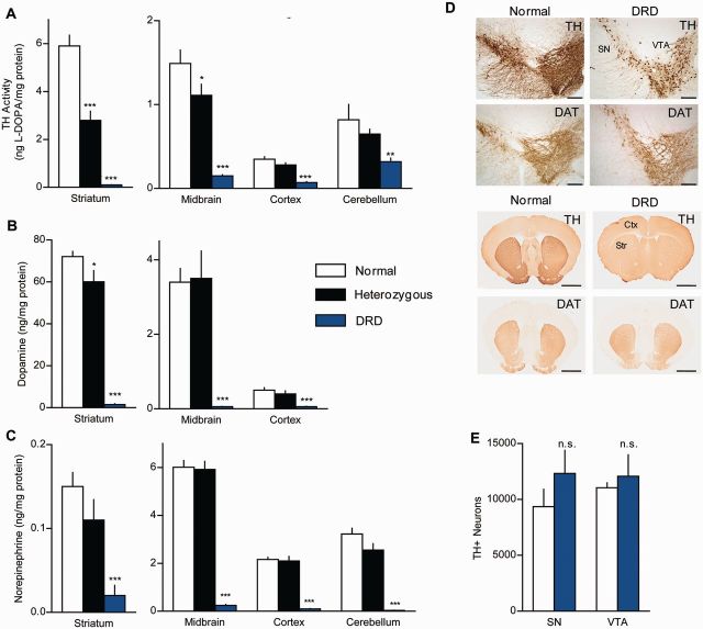
Figure 2.
TH dysfunction in DRD mice. (A) TH activity was assessed in vivo in normal (n = 5), heterozygous (n = 7), and DRD mice (n = 8). TH activity was significantly reduced in all brain regions tested in DRD mice, including striatum [F(2,17) = 96.5, P < 0.001], midbrain [F(2,17) = 44.1, P < 0.001], cortex [F(2,17 = 42.3, P < 0.001] and cerebellum [F(2,17) = 8.3, P < 0.01]. (B and C) Regional analysis of dopamine and norepinephrine concentrations in normal (n = 9), heterozygous (n = 6), and DRD mice (n = 6). Dopamine was significantly reduced in striatum [F(2,18) = 137.4, P < 0.001], midbrain [F(2,18) = 17.1, P < 0.001], and cortex [F(2,18) = 15.0, P < 0.001]. Norepinephrine was significantly reduced in striatum [F(2,18) = 10.0, P < 0.001], midbrain [F(2,18) = 136.7, P < 0.001], cortex [F(2,18) = 100.2, P < 0.001] and cerebellum [F(2,18) = 54.5, P < 0.001]. (D) Representative sections immunostained for TH or DAT from striatum or midbrain of normal and DRD mice (Scale bars = 1.5 mm, striatum; 200 µm, midbrain). (E) Stereological cell counts of TH-positive neurons in the substantia nigra (SN) (P > 0.1, Student’s t-test) and ventral tegmental area (VTA) (P > 0.1, Student’s t-test). l-DOPA supplementation was terminated >24 h before sacrificing animals for analysis at 8 pm. Values represent mean ± SEM. Statistical analyses for TH activity and monoamine concentrations were performed for each region using a one-way ANOVA with a Holm-Sidak post hoc analyses where appropriate; *P < 0.05, **P < 0.01, ***P < 0.001 compared to normal.

