Figure 3.
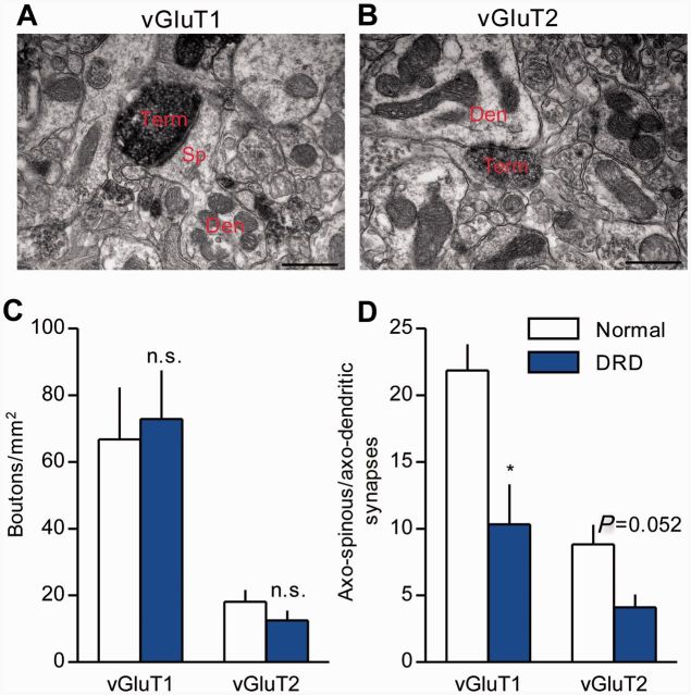
Glutamatergic cortico- and thalamo-striatal terminals in DRD mice. (A and B) Examples of vGluT1- (A, corticostriatal) or vGluT2- (B, thalamostriatal) immunoreactive terminals forming an axo-spinous (A) or an axo-dendritic (B) asymmetric synapse in the mouse dorsolateral striatum. Scale bars = 500 nm in A and B. (C) No significant difference was observed in the density of either vGluT1-positive or vGluT2-positive terminals between the genotypes (n = 3/genotype; Student’s t-test). (D) The ratio of vGluT1-positive (n = 3 animals/genotype, 177-180 terminals/genotype) or vGluT2-positive (n = 3/genotype, 133–155 terminals/genotype) axo-spinous to axo-dendritic synaptic contacts was measured. This ratio was significantly smaller (P < 0.05, Student’s t-test) for vGluT1-positive terminals, and approached significance for vGluT2-positive terminals (P = 0.052, Student’s t-test), in the dorsolateral striatum of DRD mice. Values represent mean ± SEM; *P < 0.05. Den = dendrite; Sp = spine; Term = terminal.

