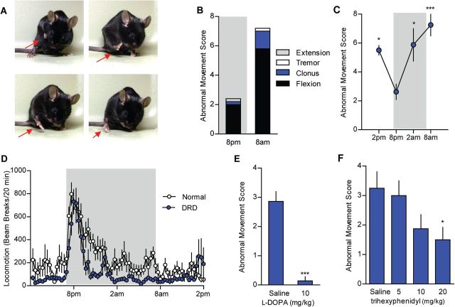
Figure 4.
The DRD behavioural phenotype. (A) DRD mice exhibit dystonic movements that occurred most often in the limbs. (B) The abnormal movements were largely composed of transiently sustained flexion in both the inactive (light) and active (dark) period. Data collected in the active are shaded in grey. (C) Dystonic movements were assessed in DRD mice over the course of a day (n = 8). DRD mice expressed significantly fewer dystonic movements at 8 pm than at other times of day [F(3,28) = 6.8, one-way repeated measures ANOVA, P < 0.05 8 pm versus 2 am and 2 pm, P < 0.001 8 pm versus 8 am, Tukey’s post hoc analysis]. (D) Spontaneous locomotor activity was assessed over 24 h in normal and DRD mice (n = 10/genotype). Locomotor activity was significantly reduced in DRD mice compared to normal mice across the entire 24 h period [F(1,18) = 9.6, two-way repeated measures ANOVA; P < 0.01, Holm-Sidak post hoc analysis]. In the active period, there was a significant genotype × time interaction effect [F(1,18) = 5.2, two-way repeated measures ANOVA, P < 0.05, Holm-Sidak post hoc analysis] reflecting the reduction in locomotor activity in DRD mice in the last 6 h of the active period. (E) Peripheral administration of l-DOPA significantly reduced dystonic movements in DRD mice (n = 7/treatment; P < 0.001, paired Student’s t-test) at 2 pm. (F) Administration of trihexyphenidyl (n = 8/dose), a muscarinic acetylcholine receptor antagonist, significantly reduced dystonic movements in DRD mice [F(3,21) = 3.4, one-way repeated measures ANOVA, P < 0.05, Holm-Sidak post hoc analysis] at 2 pm. Values represent mean ± SEM; *P < 0.05, **P < 0.01, ***P < 0.001.

