Fig. 4.

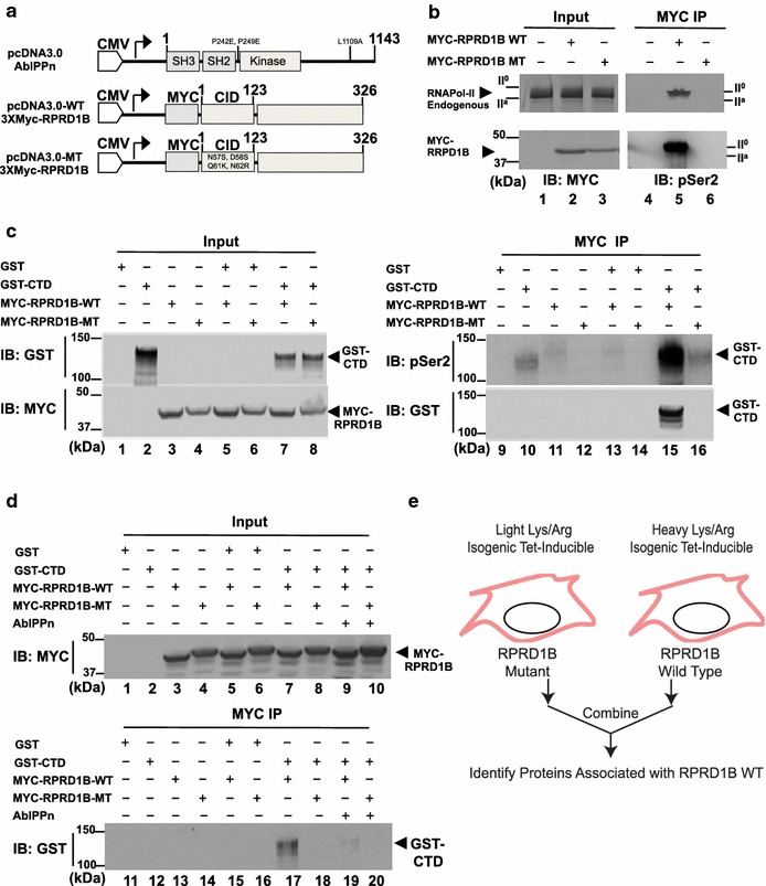
Interaction of full-length Myc-RPRD1B but not CID-mutant Myc-RPRD1B with RNAPII and the CTD. a Diagram of full-length wild type (WT) RPRD1B and mutant (MT) RPRD1B containing the following point mutations N57S, D58S, Q61K, N62R in the CID. AblPPn is a constitutively activated Abl kinase in which two proline residues (PP) in the SH2-kinase linker are mutated to glutamic acids to disrupt auto-inhibition, and a leucine residue to inactivate the nuclear export signal (n). b WT but not MT RPRD1B interacts with endogenous RNAPII. Total lysates from HEK293T cells transfected vector (lane 1), WT Myc-RPRD1B (lane 2), MT Myc-RPRD1B (lane 3), were immunoblotted with the anti-Myc and anti-8WG16 to detect RPRD1B and RNAPII (left panels). These cell lysates were also immunoprecipitated with anti-Myc (9E10) to detect the co-immunoprecipitation of endogenous RNA polymerase II (N20) and pS2-CTD (right panels). c WT but not MT RPRD1B interacts with recombinant CTD. Total lysates from HEK293T cells transfected with GST (lane 1), GST-CTD (lane 2), or combinations with Myc-RPRD1B (lanes 3–8) and probed with anti-GST or anti-Myc (left panels, input). These lysates were also immunoprecipitated with anti-Myc (9E10) and probed with anti- pSer2-CTD or anti-GST. d C-transfection with AblPPn inhibited RPRD1B interaction with CTD. Total lysates (input) from HEK293T cells transfected with GST (lane 1), CTD (lane 2), WT RPRD1B (lane 3), MT RPRD1B (lane 4), or combinations (lanes 5–8) including those with AblPPn (lanes 9, 10) were probed with anti-Myc (upper panel). These ten lysates (1–10) were immunoprecipitated with anti-Myc (MYC IP) and the immunoprecipitates probed with anti-GST (lanes 11–20, lower panel). Note that GST-CTD associated with WT but not MT RPRD1B, and that AblPPn disrupted WT RPRD1B interaction with GST-CTD (compare lane 17–19). e Diagram of SILAC mass spectrometry strategy used to identify proteins that associated with WT but not MT RPRD1B. Heavy and light isotope labeling was conducted after tetracyclin-induced expression of the WT and MT RPRD1B in HEK293T cells. See Tables 3 and 4 for summaries of SILAC results
