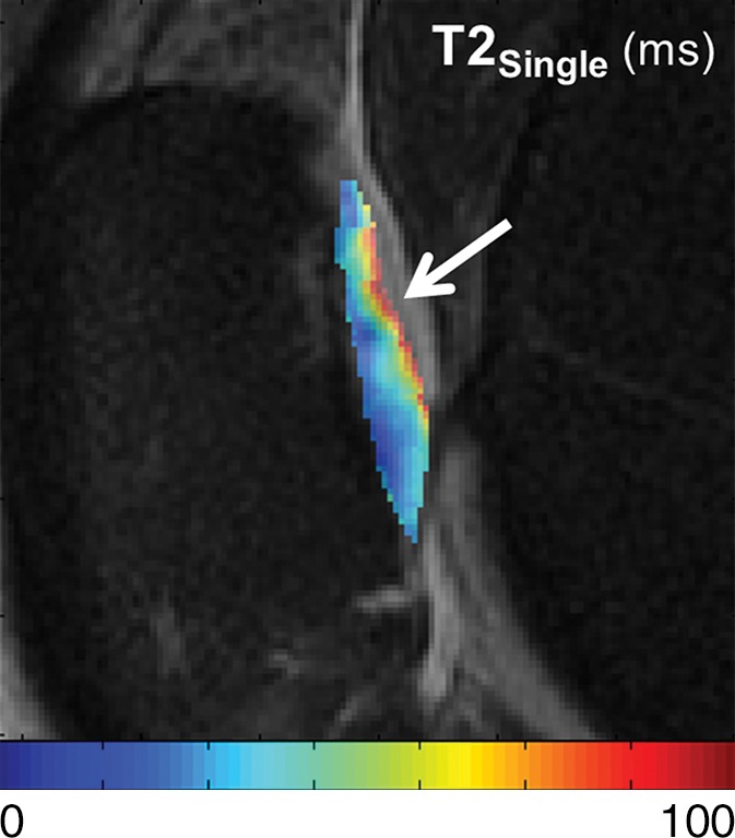
Figure 4b:

T2Single, T2F, T2S, and FF maps show articular cartilage of knee joint in a 49-year-old man with Kellgren-Lawrence grade 2 osteoarthritis of the knee. (a) 3D FSE image shows focal area of superficial partial-thickness cartilage loss on patella (arrow). Corresponding (b) T2Single, (c) T2F, (d) T2S, and (e) FF maps show increased T2Single, T2F, and T2S, and decreased FF in patellar cartilage (arrow) at the site of superficial cartilage lesion. Note that changes in FF are more pronounced and extend deeper into cartilage than other single-component and multicomponent T2 parameters.
