
Figure 5.
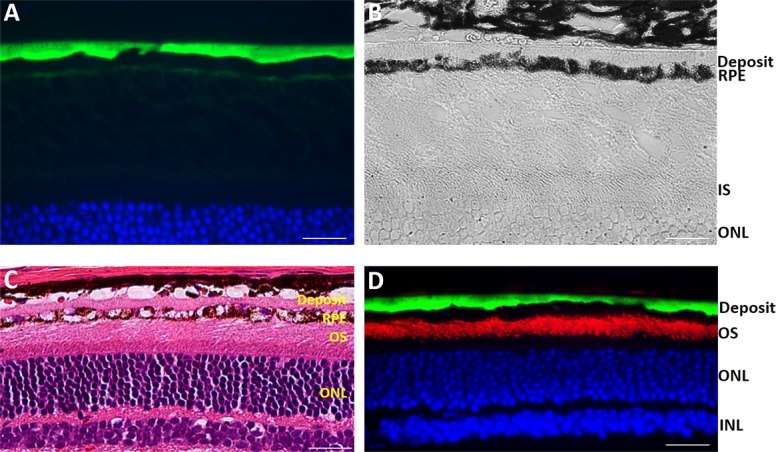
Examination of S163R expression at 9 months following AAV-hC1QTNF5(S163R)–HA delivery. (A) Detection of the AAV-expressed S163R by immunofluorescence. Note the thick, continuous S163R basal deposit, which appears transparent in the brightfield above the pigmented RPE (B). (C) Staining with H & E showing the thick basal deposit above a highly vacuolated, deteriorated RPE. (D) Double-labeling with an antibody against rod opsin showing the outer segments (red), and anti-HA (green) detecting the thick S163R deposit basal to the RPE. Scale bars: 20 μm.