
Figure 6.
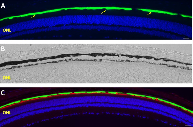
Widespread distribution of basal S163R deposits at 9 months following AAV-hC1QTNF5(S163R)–HA delivery. (A–C) Low magnification images of retinal sections showing continuous, widespread S163R expression (green) above the RPE by immunofluorescence (A, C). (B) Brightfield image showing the transparent basal S163R deposit between the choroid and the pigmented RPE. (C) The RPE apical membrane is shown in red as labeled by the anti-MFRP antibody.