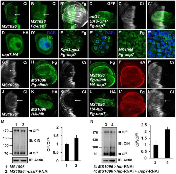
Figure 3. Usp7 Counteracts Ci Degradation Mediated by Both Slimb-Cul1 and Hib-Cul3 E3 ligases.
UAS transgene expression with MS1096 gal4 is usually stronger in the dorsal region than in the ventral of wing pouch.
(A) A wing disc of MS1096 line was stained to show full-length Ci (green).
(B-B’) A wing discs expressing Fg-usp7 with MS1096 was immunostained with Fg (white) and Ci (green) antibodies. Ci was accumulated when usp7 was overexpressed (arrow).
(C-C”) A wing disc expressing Fg-usp7 with apG4 was immunostained to show the expression of GFP (green) and Ci (white).
(D-D’) Expressed Usp7-HA (green) in S2 cells mainly localized in nuclei.
(E-E’) Fg-Usp7 protein (green) expressed by sgs3-gal4 in salivary glands mainly localized in nuclei.
(F-F’) Fg-Usp7 protein (green) expressed by MS1096 in wing discs mainly localized in nuclei. From D to F’, the nuclei were marked by DAPI (blue)
(G-I”) A MS1096 wing disc (G) and the discs expressing Fg-slimb alone (H-H’) or Fg-slimb/HA-usp7 together (I-I”) by MS1096 were immunostained to show Fg tag (green), Ci (white), and HA tag (red). Usp7 could counteract Ci degradation by Slimb-Cul1 E3 ligase (arrows).
(J-L”) A MS1096 wing disc (J) and the discs expressing HA-hib alone (K-K’) or HA-hib/Fg-usp7 (L-L”) together by MS1096 were immunostained with HA tag (green), Ci (white), and Fg tag (red) antibodies. Usp7 could attenuate Ci degradation by Hib-Cul3 E3 ligase (arrows).
(M-N) Western blot analysis of lysates from control wing discs or wing discs expressing indicated RNAi using MS1096 driver. Approximately 40 discs were dissected, lysed and blotted with rabbit anti-CiN antibody, respectively. Intensity ratio of CiR to CiFL of each lane was shown on the right. The ratio result was presented as means±SD of values from three independent experiments.
See also Figure S4.

