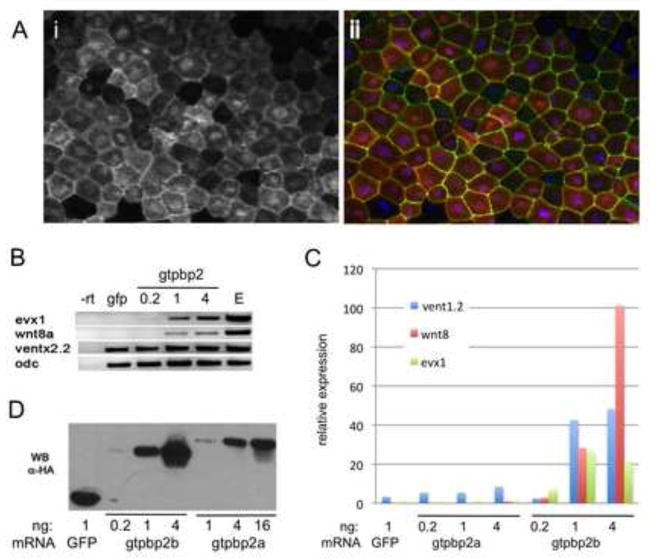
Figure 6. Gtpbp2 subcellular localization and induction of BMP target genes.
A. mCherry-tagged Xenopus gtpbp2a mRNA (2ng) co-injected with a membrane-localized GFP (mem-GFP) mRNA (10 pg), and costained with DAPI, shows nuclear and cytoplasmic localization in gastrula stage 11 animal cap cells; (i) mCherry-Gtpbp2 signal only, (ii) merged signals for mCherry-Gtpbp2b, mem-GFP and DAPI. B. Xenopus laevis Gtpbp2b can induce BMP marker genes evx1, wnt8a, vent2.2 in animal caps. gtpbp2b mRNA was injected at increasing doses (0.2ng, 1ng, and 4ng) into the animal pole of two cell stage embryos, animal caps were excised at stage 8, and cultured to stage 11 before gel-based RT-PCR with primers to indicated genes (otx as positive control for RT and loading). C. Q-PCR analysis of the bmp targets vent1.2, wnt8, and evx1 in animal caps injected at the 2 cell stage with increasing doses of gtpbp2a or gtpbp2b mRNA, excised at the 8 cell stage, and cultured to stage 11. D. Comparison of accumulation of HA-tagged Xenopus laevis Gtpbp2a versus Gtpbp2b isoforms in embryos injected with increasing doses of mRNA at the 2 cell stage and lysed at stage 11 for western blotting using an anti-HA antibody.

