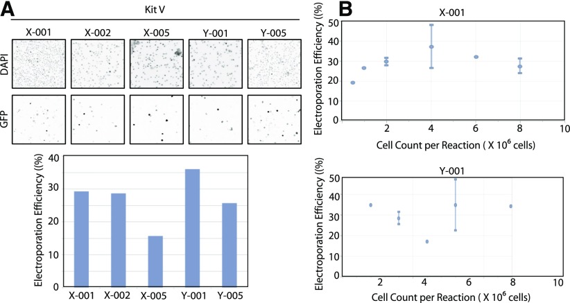
Figure 1.
Optimization of electroporation of GM12878 cell transfection efficiency by use of GFP-expressing plasmids. A) GM12878 cells (2 × 10⁶) were transfected with GFP-expressing plasmid by use of Lonza Kit V with the indicated channels. Twenty-four hours post-transfection, the cells were stained with Hoechst nuclear DNA stain (1 μg/ml) for 1 h. All cells were imaged with an inverted fluorescence microscope (upper), and the percentage of GFP-expressing cells was quantitated (lower). Images shown in A have been inverted for easier viewing. B) GM12878 cells at varying cell densities were transfected with 2 μg GFP-expressing plasmid by use of Lonza Kit V with the indicated channels. Twenty-four hours post-transfection, the cells were stained with Hoechst nuclear DNA stain (1 μg/ml) for 1 h. The percentage of GFP-expressing cells was quantified by use of the ImageJ software program (NIH).

