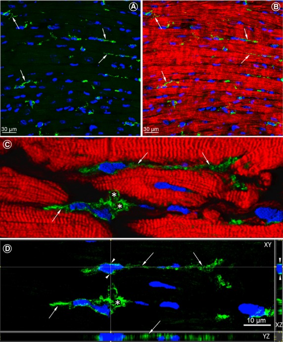
Figure 1.

Confocal images of c-kit-positive telocytes (TCs) in longitudinal sections of the normal human LV myocardium. (A) Numerous c-kit-positive cells (green) with long cytoplasmic processes are indicated by arrows. (B) Merged view of the image shown in A showing the spatial relationship of TCs with the neighbouring cardiomyocytes stained red with Alexa633-conjugated phalloidin. (C) Two TCs located in close vicinity with cardiomyocytes. Arrows denote telopodes (Tps) originating from the cell body. Asterisks indicate labyrinthine systems. (D) Identical images as in B viewed in XY, XZ and YZ axes. Arrowhead points the very small rim of the perinuclear cytoplasm. Thin and long TPs are indicated with arrows and the asterisk indicates the labyrinthine system. Nuclei are stained blue with 4,6-diamidino-2-phenylindole (DAPI).
