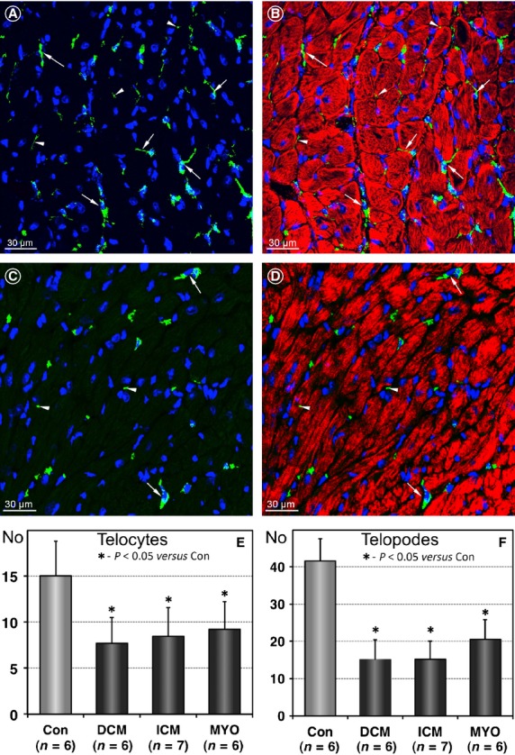
Figure 2.

Telocytes (TCs) and telopodes (Tps) are reduced in diseased human hearts. (A and B) Representative immunofluorescent confocal images showing the distribution of c-kit-positive cells (green) in a normal heart. Arrows denote TCs and arrowheads indicate TPs. (C and D) Confocal images of TCs in a patient with DCM demonstrating fewer TCs (arrows) and TPs (arrowheads) than in controls. Cardiomyocytes are stained red with Alexa633-conjugated-phalloidin. Nuclei are stained blue with 4,6-diamidino-2-phenylindole (DAPI). (E and F) Results of quantification of the number of TCs and TPs per 1 mm2 myocardial area in controls and heart failure patients.
